Our Research
Decoding the hidden communication systems that allow life to persist across generations.
Sexually reproducing organisms must carefully break, repair, and reorganize their DNA to generate healthy, genetically diverse offspring. The Diezmann Lab studies how chromosomes coordinate these transformations during meiosis, investigating the hidden signaling systems that allow cells to preserve genetic integrity while reshaping it across generations.
To study these processes, we combine genetics, live-cell microscopy, and quantitative biophysics to observe molecular interactions inside living cells. Using single-molecule imaging in the model organism C. elegans, we track how proteins organize along chromosomes, revealing the dynamic physical systems that guide DNA repair and recombination.
By uncovering the fundamental rules that govern molecular coordination, our work advances understanding of fertility, DNA repair, evolution, and the self-organization of living systems. These discoveries may ultimately inform new strategies in human medicine, while deepening our understanding of how life maintains and transforms itself over time.
Major Themes in Our Work
-
![]()
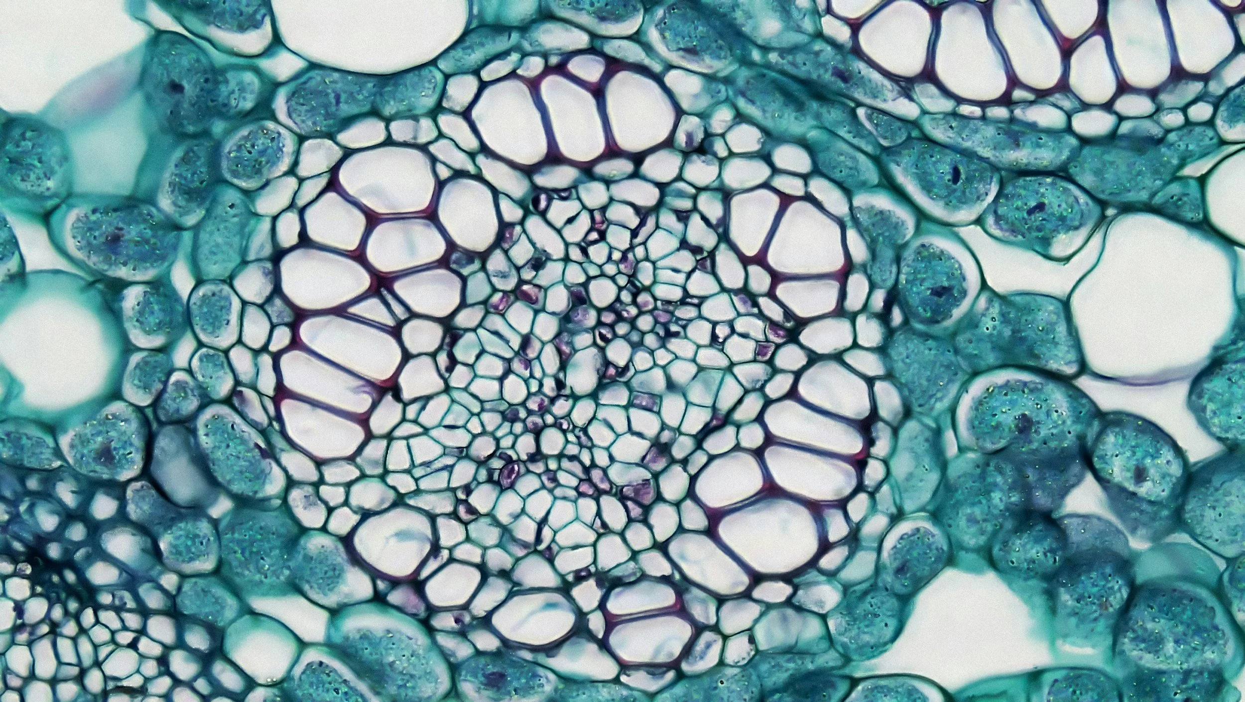
Condensates, Flux, and Interfaces
We study how molecules come together and communicate across differences, from the nanoscale to the cell scale.
-
![]()
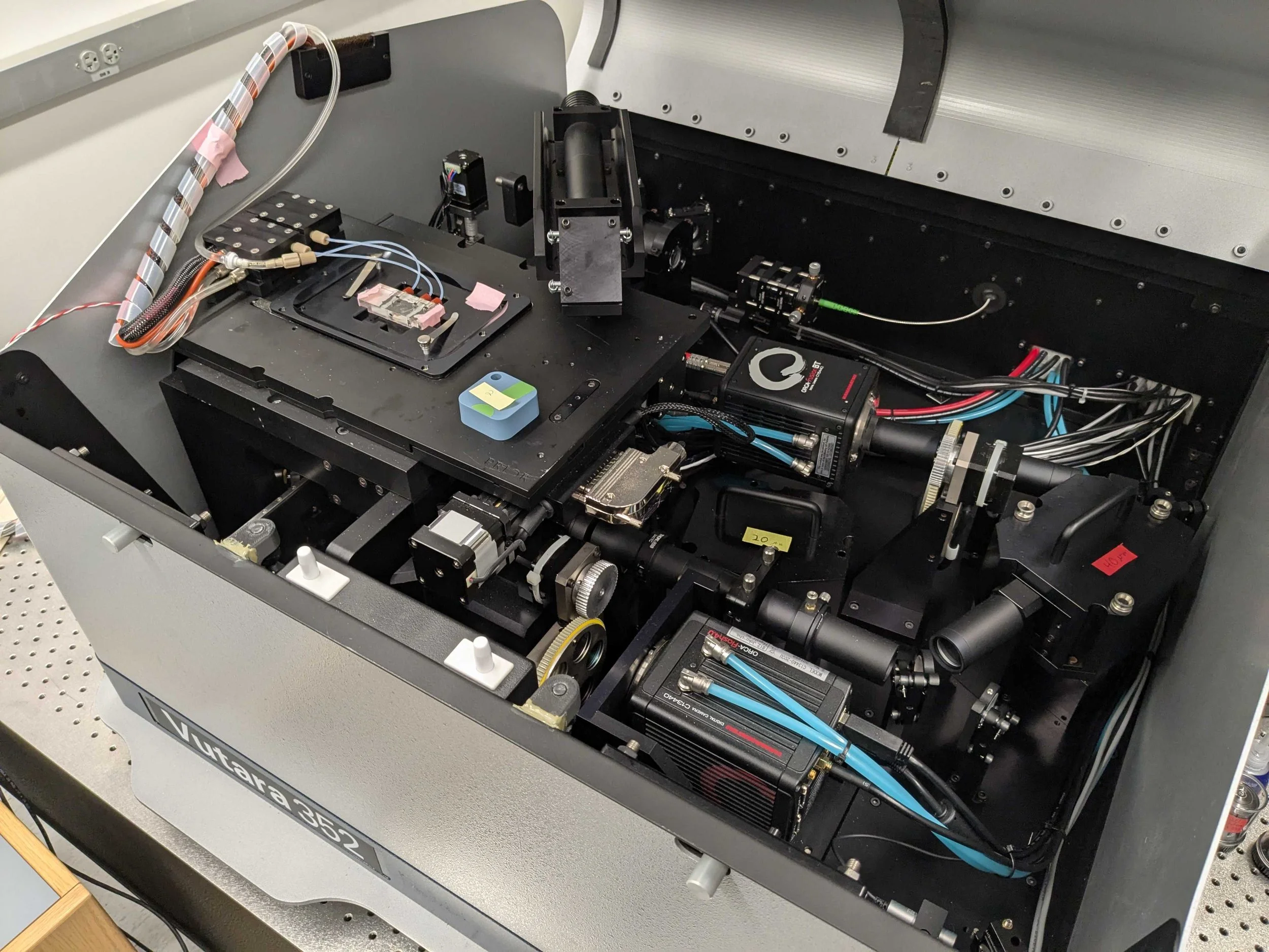
Precision Microscopy and Genetics
We use tools that let us see and follow molecular dynamics, and make precise edits to molecular function.
-
![]()
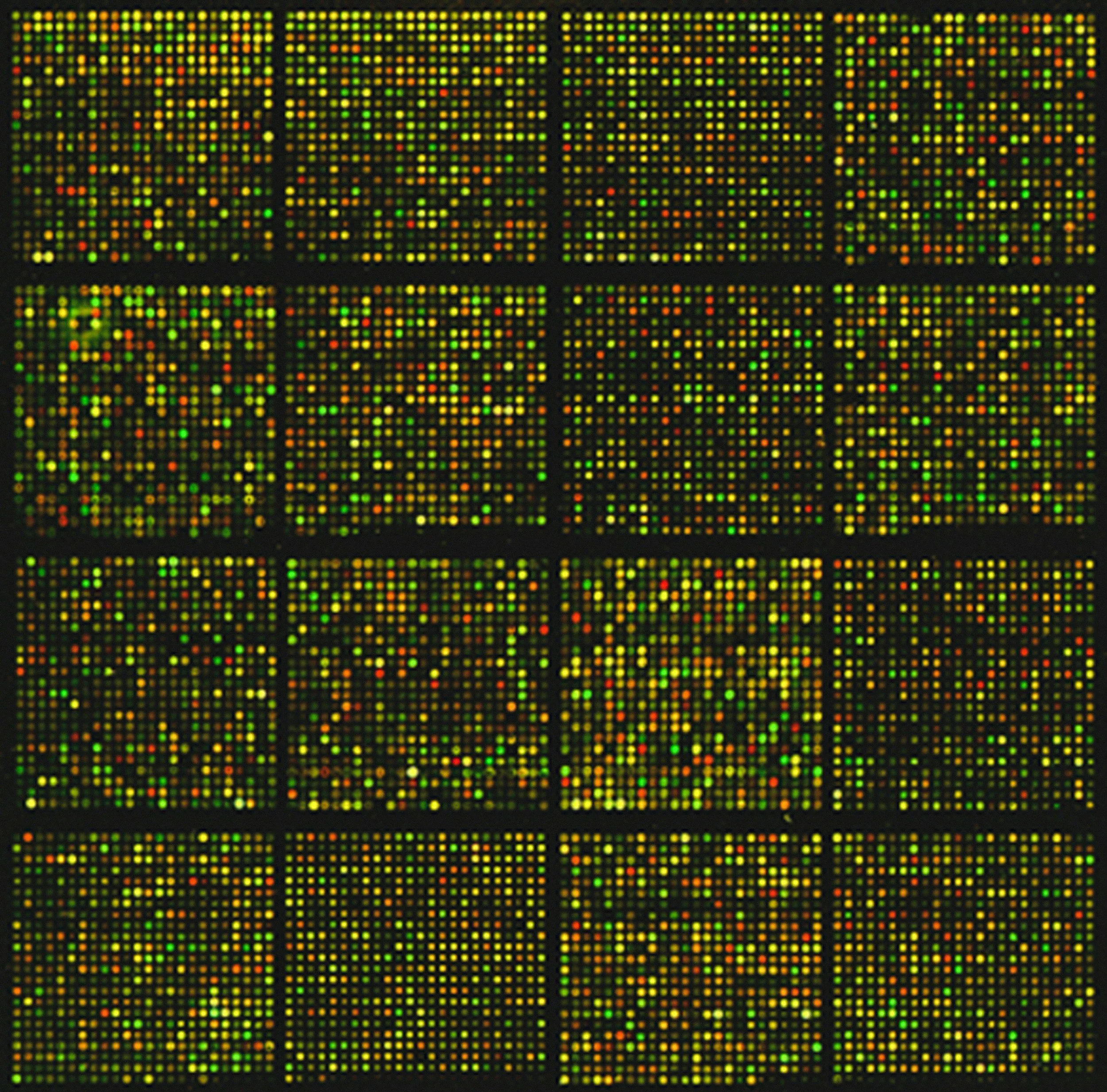
Symmetry-Breaking Decisions
We want to know how cells integrate complex information to select between possible outcomes - for instance, in DNA repair.

Featured Project
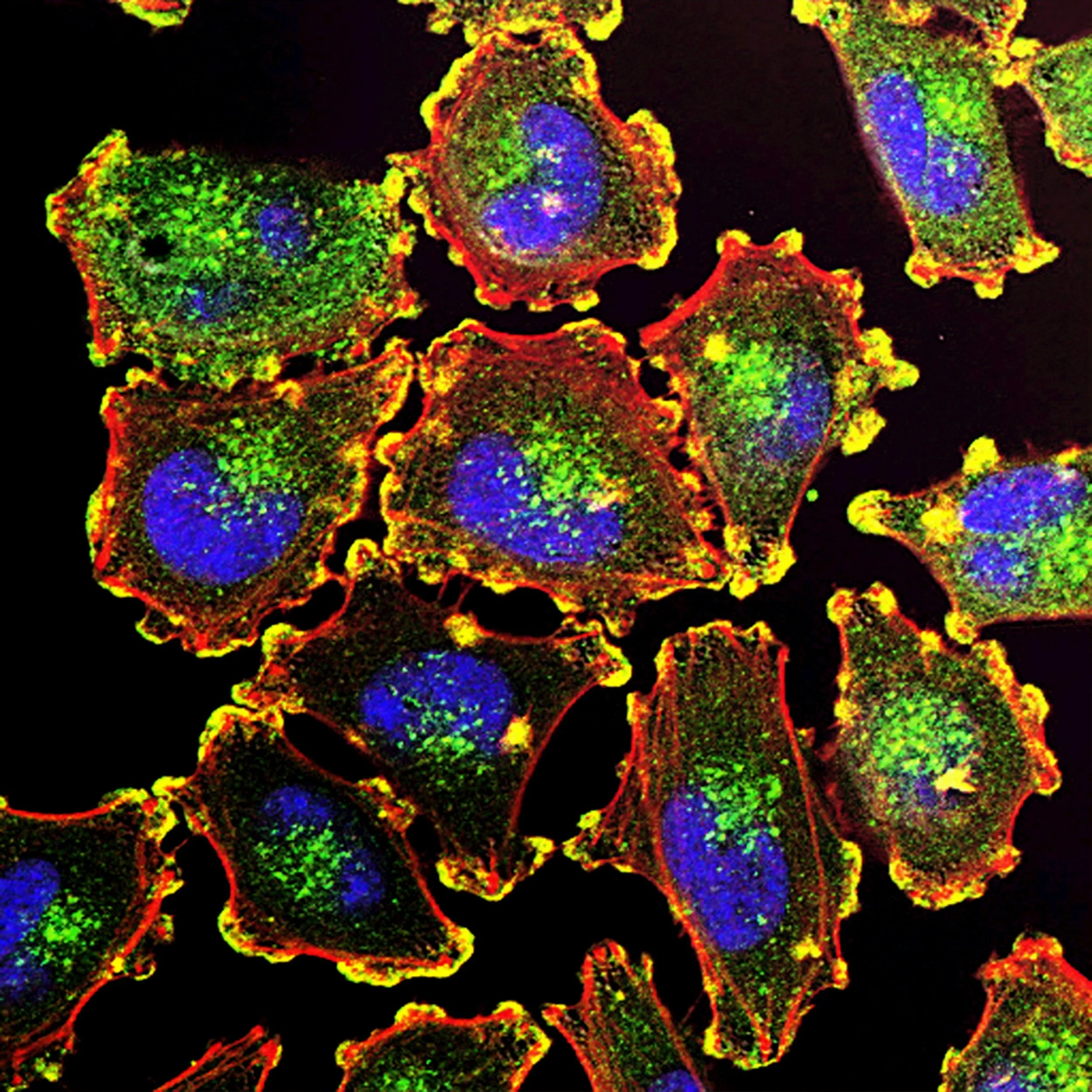
Single-Molecule Tracking Reveals Self-Organization At The Nanoscale
We study single-molecule microscopy: A technology that watches individual molecules moving within complex environments, at the nanoscale. It works by imaging single fluorescent molecules. Fluorescent molecules are tagged to let us image them inside cells. By blinking them on and off randomly, we can see them one at a time. By localizing them to nanoscale precision, we can generate nanoscale pictures of where molecules are, or we can even follow them in real time as they explore cells.
Incredibly, this technique can be applied to complex biological molecules, like proteins, within living cells. By watching how proteins explored apparently simple bacterial cells, we learned that protein self-organization creates "sticky" interactions between proteins at distinct locations within the cells. This explains how even simple cells divide asymmetrically, a fundamental mystery related to how our own stem cells differentiate.
Explore our Publications: Optica | Chemical Review



Featured Project
Direct Imaging of Meiotic Signaling Proteins Moving Along Chromosomes
I then turned to a different kind of fate decision: How our bodies develop gametes that combine genetic material from both parents.
It turns out that this question also begins at the nanoscale: The thin, 100-nm thick ribbon that forms between paired parental chromosomes during egg or sperm development. Critically, this protein interface plays a key role in defining the sites of "crossing over" where DNA repair is used to cut and paste our parents' chromosomes together in new ways. Applying our single-molecule perspective to this system, I found that the signals that define crossovers diffuse along chromosomes using the SC as a conduit, supporting an exciting new possible mechanism for this process.

Featured Project
Material Properties Regulate Binding and Diffusion In The SC
In the lab, we have been pushing the boundaries of this technique by beginning to look at the impact of conditions like temperature and protein-protein interactions of dynamics of scaffold and signaling proteins along chromosomes.
Forthcoming publications: Preparation of C. elegans Gonad Explants for High-Resolution Imaging of Living Meiotic Nuclei; Regulation of SC Properties by Temperature and Binding Interactions
Our work has been featured in